|
|
|
 |
CDC Dental Diagrams |  |
|
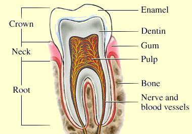
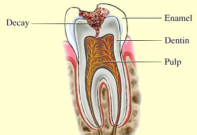
Diagrams of Tooth Structure In Health & Disease Teeth are composed of several layers including hard and soft tissues. The most internal, and most sensitive, is the Pulp tissue, where the nerves & blood vessels lie. The outermost layer is the hardest, and the least sensitive. It is the Enamel; a solid shiny tissue. The intermediate layer is the Dentine which forms the bulk of the tooth. Its properies are also intermediate between the other two layers. Tooth decay starts at the surface and invades the internal structures. In the early stages no or little pain is felt. When decay reaches the pulp pain is usually severe and the tooth may die. Pain is a late sign of dental decay, so early treatment is the best way to save a tooth. |
|
|
|
|
| Tooth Decay Decay starts at the surface and invades internal structurs. |
 |
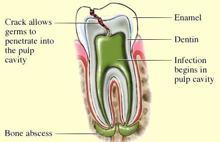 |
Formation of dental abscess A dental abscess forms as decay reaches the pulp. |
| |
|
|
|
|
| |
|
|
|
|
| |
|
|
|
|
| |
|
|
|
|
| |
|
|
|
|
| |
|